
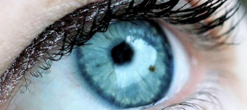
What is the macula?
The macula is an anatomical area of the retina, which is located at the eye fundus. In the macula there is a high concentration of specialized cells – photoreceptors, where the beam of light passing at any given time from the outside environment to the inside of the eye, is focused upon. The macula contains some specific photosensitive dyes, called xanthophylls, assisting in the absorption of light and protecting the macula from oxidative stress. It is responsible for sharp vision and color perception.
Classification/Treatment
The macula may be damaged by various conditions and diseases. There are two major forms of macular degeneration, atrophic and exudative. In the first case the degeneration mechanism involves the development of atrophy either of the photoreceptors, or of the pigment epithelium, the substrate layer underneath the retina and macula. The exudative macular degenerations are distinguished by the presence of liquid / edema in the retina layers.
The most common degenerations of the macula are outlined below:
1. Age-related: is an old age disease (over 50 years), where degenerative material accumulations (drusen) are generated around and into the macula. It is distinguished into dry and wet form: In the first case monitoring and taking vitamin supplements are recommended. In the second case that is further complicated by the development of abnormal vessels therapy consists of intravitreal injections of anti-angiogenic agents. A special category is the choroidal polyps requiring a special laser treatment with intravenous infusion of photosensitizing substance (photodynamic therapy).
2. Diabetic maculopathy: This is the development of edema in the macula, resulting from lesions of the retina vessels due to diabetes. The so-called clinically significant edema is treated with a combination of laser and intravitreous injections. Lately, a new technology has been developed, subthreshold diode micropulse laser for photoexcitation of the macula without burns.
3. Macular edema after venous occlusion: The central retinal vein or its branches can become occluded by various causes. The occlusion creates hemodynamic disturbances in the circulation of the retina, resulting in hemorrhages and macular edema. It is treated with intravitreal injections, cortisone implants and sometimes subthreshold diode micropulse laser.
4. Central serous chorioretinopathy: It is a degeneration usually occurring at a young age, but not exclusively. It is associated with anxiety and the macula is affected due to the presence of serous fluid in the retina. It usually automatically recedes within 3-6 months but may recur or acquire a chronic nature. In such cases, appropriate photodynamic therapy or subthreshold diode micropulse laser are indicated.
5. Dystrophies, such as Stargardt disease or Best disease: These are inherited macular diseases, wherein some abnormal genes are responsible for the early, usually in a young age, damage of the macula, which is of an atrophic type. There is no standard treatment, while in some cases a genetic check is recommended to detect the responsible gene and estimate the probability of inheritance in progeny.
Diagnosis
Besides fundoscopy, a number of specific imaging examinations are used for the diagnosis of macula degenerations, such as:
- Optical Coherence Tomography (OCT), scanning the macula in thin sections, wherein all the retinal layers in the macular region are distinct.
- Digital fluoroscopy, using a contrast medium by intravenous injection, for the imaging of retinal vessels. It is particularly indicated in exudative MD.
- Autofluorescence, with specialized capturing of the posterior fundus pole, depicting the retinal pigment epithelium. It is particularly useful in macular dystrophies, but also in inflammatory diseases of the fundus.
- Indocyanine green angiography with intravenous injection of contrast substance. Mainly imaging the choroid, the deep retinal layer.
- Electrophysiology, where the electrical activity of macular photoreceptors is recorded. It is a necessary examination for macular dystrophies.
- Ultrasound, for the diagnosis of diseases not clearly visible clinically (e.g. neoplasma of the eye).